Home » Without Label » Anatomy Rib Cage : Anatomy Of Rib Cage The Ribs Rib Cage Articulations Fracture Teachmeanatomy Wayne Vogl And Adam W M Roda Dunia : Check out our anatomy rib cage selection for the very best in unique or custom, handmade pieces from our shops.
Anatomy Rib Cage : Anatomy Of Rib Cage The Ribs Rib Cage Articulations Fracture Teachmeanatomy Wayne Vogl And Adam W M Roda Dunia : Check out our anatomy rib cage selection for the very best in unique or custom, handmade pieces from our shops.
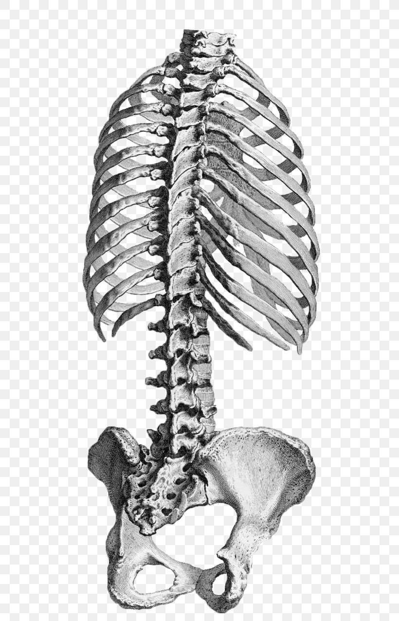
Anatomy Rib Cage : Anatomy Of Rib Cage The Ribs Rib Cage Articulations Fracture Teachmeanatomy Wayne Vogl And Adam W M Roda Dunia : Check out our anatomy rib cage selection for the very best in unique or custom, handmade pieces from our shops.. Contributing to their role in protecting the internal thoracic organs. Human muscles · april 17, 2020. The thoracic cage consists of the 12 thoracic vertebrae, the associated intervertebral discs, 12 pairs of ribs with their costal cartilages, and the sternum. Each pair is numbered based on their attachment to the sternum, a bony process at the front of the rib cage which serves as an anchor point. The thoracic cage protects the heart and lungs.
The top edge of the manubrium has a depression called the suprasternal or jugular notch. Each pair is numbered based on their attachment to the sternum, a bony process at the front of the rib cage which serves as an anchor point. The cartilage strips are called costal cartilage (costal is the anatomical adjective that refers to the rib) and connect on their other end to the sternum. The rib below that is rib 2, and it connects to the t2 thoracic vertebra, and so on. Human muscles · april 17, 2020.
The Thoracic Cage The Ribs And Sternum Human Anatomy And Physiology Lab Bsb 141 from s3-us-west-2.amazonaws.com The upper edge is round and the lower sharp. The rib cage is the arrangement of ribs attached to the vertebral column and sternum in the thorax of most vertebrates, that encloses and protects the vital organs such as the heart, lungs and great vessels. Contributing to their role in protecting the internal thoracic organs. On the interior wall of the rib body is a channel, sulcus costae, with blood vessels and nerves. It is made up of 12 pairs of ribs. It consists of the 12 pairs of ribs with their costal cartilages and the sternum (figure 6.38). The rib cage shields the heart and lungs from damage. An enlarged or ruptured spleen can cause sudden or chronic pain under the left rib cage that ends up migrating towards the back and/or shoulders.
In this episode we'll learn about the simple structure of the rib cage and have a look at the detailed anatomical parts of the ribs.
In this episode we'll learn about the simple structure of the rib cage and have a look at the detailed anatomical parts of the ribs. Rib cage anatomy the rib cage, shaped in a mild cone shape and more flexible than most bone sets, is made up of varying elements such as the thoracic vertebra, 12 equally paired ribs, costal cartilage, and held together anteriorly by the sternum. The ribs are a set of twelve paired bones which form the protective 'cage' of the thorax. Contributing to their role in protecting the internal thoracic organs. Anatomy the rib cage is a bony structure found in the chest (thoracic cavity). It forms the bony framework for breathing. On the interior wall of the rib body is a channel, sulcus costae, with blood vessels and nerves. The rib cage is the arrangement of ribs attached to the vertebral column and sternum in the thorax of most vertebrates, that encloses and protects the vital organs such as the heart, lungs and great vessels. Structure of the ribcage and ribs. A rib has a flat body, as you can see from the picture of the anatomy of the human rib cage. The top edge of the manubrium has a depression called the suprasternal or jugular notch. It may occur after an obvious injury or without explanation. Ten of the twelve ribs connect to strips of hyaline cartilage on the anterior side of the body.
The upper edge is round and the lower sharp. In this video, we explore:1) the anatomy of the sternum2) the anatomy and differences between the three classes of ribs3) the anatomy and differences between. The bones of the rib cage are the sternum, the 12 thoracic vertebrae and the 12 pairs of ribs. It is made up of 12 pairs of ribs. Check out our anatomy rib cage selection for the very best in unique or custom, handmade pieces from our shops.
Ribcage Musculoskeletal Key from i1.wp.com The thoracic cage consists of the 12 thoracic vertebrae, the associated intervertebral discs, 12 pairs of ribs with their costal cartilages, and the sternum. Human muscles · april 17, 2020. The top edge of the manubrium has a depression called the suprasternal or jugular notch. There are twelve pairs of ribs, all of which articulate with the vertebral column. The sternum is a flat bone that is made up of three parts, the (1) manubrium, (2) body, and the (3) xiphoid process. Rib cage pain may be sharp, dull, or achy and felt at or below the chest or above the navel on either side. This furrow isn't present in the 11th and 12th ribs. Ten of the twelve ribs connect to strips of hyaline cartilage on the anterior side of the body.
Shadows around the rib cage (eg, rib companion shadows, sharp lines along the lower margin of the ribs, rib overlying shadows) may mimic pleural and extrapleural disease on frontal chest radiographs.
The top edge of the manubrium has a depression called the suprasternal or jugular notch. Check out our anatomy rib cage selection for the very best in unique or custom, handmade pieces from our shops. Rib cage anatomy the rib cage, shaped in a mild cone shape and more flexible than most bone sets, is made up of varying elements such as the thoracic vertebra, 12 equally paired ribs, costal cartilage, and held together anteriorly by the sternum. Ten of the twelve ribs connect to strips of hyaline cartilage on the anterior side of the body. The bones of the rib cage are the sternum, the 12 thoracic vertebrae and the 12 pairs of ribs. The ribs are a set of twelve paired bones which form the protective 'cage' of the thorax. A rib has a flat body, as you can see from the picture of the anatomy of the human rib cage. At the chest, many rib bones connect to the sternum via costal cartilage,. Rib cage, in vertebrate anatomy, basketlike skeletal structure that forms the chest, or thorax, and is made up of the ribs and their corresponding attachments to the sternum (breastbone) and the vertebral column. In spite of its resistance, the cage is dynamic, allowing pulmonary ventilation to. The thoracic cage protects the heart and lungs. The upper edge is round and the lower sharp. Human rib cage anatomy 3d model.
The dome shaped thoracic cage provides the necessary rigidity for organ protection, weight support for the upper limbs and anchorage for muscles. Quads only geometries (no tris/ngons). Rib cage pain can be caused. It may occur after an obvious injury or without explanation. In spite of its resistance, the cage is dynamic, allowing pulmonary ventilation to.
Human Anatomy Rib Cage Vertebral Column Pelvis Png 626x1276px Anatomy Atlas Black And White Bone Drawing from img.favpng.com Human rib cage anatomy 3d model. It is made up of 12 pairs of ribs. They articulate with the vertebral column posteriorly, and terminate anteriorly as cartilage (known as costal cartilage). Shadows around the rib cage (eg, rib companion shadows, sharp lines along the lower margin of the ribs, rib overlying shadows) may mimic pleural and extrapleural disease on frontal chest radiographs. Contributing to their role in protecting the internal thoracic organs. The spleen is used to filter red blood cells and hangs in the upper part of the abdomen. Anatomy the rib cage is a bony structure found in the chest (thoracic cavity). The top edge of the manubrium has a depression called the suprasternal or jugular notch.
Each pair is numbered based on their attachment to the sternum, a bony process at the front of the rib cage which serves as an anchor point.
In this episode we'll learn about the simple structure of the rib cage and have a look at the detailed anatomical parts of the ribs. Diagram of human body, liver rib cage, rib cage diagram labeled, rib cage diagram numbered, rib cage diaphragm, rib cage heart, rib cage organs anatomy, rib cage pain, stomach, diagram of human body, liver rib cage, rib cage diagram labeled, rib cage diagram numbered, rib cage diaphragm, rib cage. 16 photos of the rib cage diagram with organs. Instead, anatomists classify the ribs as flat bones, and they are located within the axial skeleton. The sternum is a flat bone that is made up of three parts, the (1) manubrium, (2) body, and the (3) xiphoid process. The thoracic cage (rib cage) forms the thorax (chest) portion of the body. Check out our anatomy rib cage selection for the very best in unique or custom, handmade pieces from our shops. The top edge of the manubrium has a depression called the suprasternal or jugular notch. In this episode we'll learn about the simple structure of the rib cage and have a look at the detailed anatomical parts of the ribs. Shadows around the rib cage (eg, rib companion shadows, sharp lines along the lower margin of the ribs, rib overlying shadows) may mimic pleural and extrapleural disease on frontal chest radiographs. Contributing to their role in protecting the internal thoracic organs. At the chest, many rib bones connect to the sternum via costal cartilage,. This furrow isn't present in the 11th and 12th ribs.